Back Muscles Anatomy Chart / The Female Muscular System Laminated Anatomy Chart : The superficial back muscles are the muscles found just under the skin.
Back Muscles Anatomy Chart / The Female Muscular System Laminated Anatomy Chart : The superficial back muscles are the muscles found just under the skin.. A basic understanding of the anatomy of your lower back can help you identify and differentiate a problem that commonly affects this region, such as localized muscle pain or sciatica. They also attach your shoulders and pelvis to the trunk, creating a bridge between. This article looks at the anatomy of the back, including bones, muscles, and nerves. Related posts of back muscles chart muscle anatomy trivia. Some of the links in the post above are affiliate links..
We are pleased to provide you with the picture named anatomy of back muscles diagram. Related posts of back muscles chart muscle anatomy trivia. Another common cause of lower back and hip pain is disc injury. It also covers some common conditions and injuries that can affect the back. This rotator cuff muscle helps with the raising and lowering of the upper arm.;
Labeled Anatomy Chart Of Male Triceps And Back Muscles On White Background Stock Photo Download Image Now Istock from media.istockphoto.com Back muscles, back muscle diagram. The intermediate layer contains the erector spinae muscles, whose many functions include the extension and lateral flexion of the spine, head and neck. All about the back muscles the back anatomy includes the latissimus dorsi, trapezius, erector spinae, rhomboid, and the teres major. Other muscles are small and cover much less space. To learn more about the anatomy of the spine, watch this video. This diagram depicts muscles of the back diagram.human anatomy diagrams show internal organs, cells, systems, conditions, symptoms and sickness information and/or tips for healthy living. Creatine research more than a sports supplement read more…. Superficial, intermediate, deep and deepest layers.these muscles lie on each side of the vertebral column, deep to the thoracolumbar fascia they span the entire length of the vertebral column, extending from the cranium to the pelvis
Spinal vertebrae bone spine vertebra toracica spinal cord spine structure back diagram spine sections spinal cord vertebrae spinal structure health diagram.
Superficial back muscles, intermediate back muscles and intrinsic back muscles.the intrinsic muscles are named as such because their embryological development begins in the back, oppose to the superficial and intermediate back muscles which develop elsewhere and are therefore classed as extrinsic muscles. This diagram depicts muscles of the back diagram.human anatomy diagrams show internal organs, cells, systems, conditions, symptoms and sickness information and/or tips for healthy living. Muscle origin insertion action innervation artery notes; However, the spinal erectors travel the length of the entire spine. Most of the time, back muscle pain is diagnosed then treated with little more than a prescription of rest, painkillers and muscle relaxants. Leaning back to straight vertical and all points in between. Related posts of back muscles chart muscle anatomy trivia. Lower back muscle diagram anatomy does degenerative disc disease affect the lower back muscle? 149 best skeletal muscle images in 2017 muscle tissue. Creatine research more than a sports supplement read more…. Deep back muscles diagram the superficial layer contains the splenius cervicis and splenius capitis muscles. Mastoid process and lateral end of the superior nuchal line: The back's muscles start at the top of the back (named the cervical vertebrae) and go to the tailbone (also named the coccyx).
Deep back muscles diagram the superficial layer contains the splenius cervicis and splenius capitis muscles. Leaning back to straight vertical and all points in between. Your clients will thank you for it! They extend and rotate the head and neck. This diagram depicts anatomy of back muscles.human anatomy diagrams show internal organs, cells, systems, conditions, symptoms and sickness information and/or tips for healthy living.
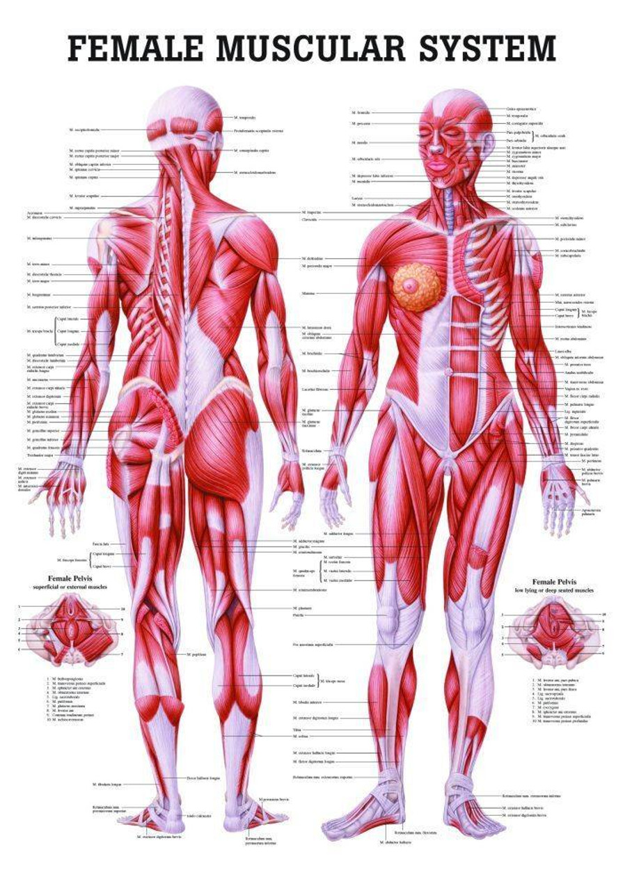
The Female Muscular System Laminated Anatomy Chart from cdn11.bigcommerce.com Muscles found in the superficial group include rhomboid major, rhomboid minor, levator scapulae, trapezius, latissimus dorsi. Spinal vertebrae bone spine vertebra toracica spinal cord spine structure back diagram spine sections spinal cord vertebrae spinal structure health diagram. Superficial back muscles, intermediate back muscles and intrinsic back muscles.the intrinsic muscles are named as such because their embryological development begins in the back, oppose to the superficial and intermediate back muscles which develop elsewhere and are therefore classed as extrinsic muscles. Muscle charts of the human body. 149 best skeletal muscle images in 2017 muscle tissue. Link to client back care guide. Leaning back to straight vertical and all points in between. Other muscles are small and cover much less space.
There are three different muscle groups found in the back:
Back muscles, back muscle diagram. This diagram depicts muscles of the back diagram.human anatomy diagrams show internal organs, cells, systems, conditions, symptoms and sickness information and/or tips for healthy living. Certain back muscles extend to other areas, like the shoulders, upper arms, and thighs. 12 photos of the muscles of the lower back and hip diagram. Another common cause of lower back and hip pain is disc injury. Anatomy chart courtesy of fcit the latissimus dorsi muscles (also known as the lats) are the largest muscles of the back. They also attach your shoulders and pelvis to the trunk, creating a bridge between. In this image, you will find 1st cervical vertebrae, atlus, cervical plexus, 7th cervical vertebrae, 1st thoracic vertebrae, brachial plexus, spinal dura mater, filaments of spinal nerve roots, 12th thoracic vertebra, 1st lumber vertebra, iliohypogastric nerve, ilioinguinal nerve, lumbar. Spinal vertebrae bone spine vertebra toracica spinal cord spine structure back diagram spine sections spinal cord vertebrae spinal structure health diagram. It also helps in extension and lateral flexion of the lumbar spine. This article looks at the anatomy of the back, including bones, muscles, and nerves. All about the back muscles the back anatomy includes the latissimus dorsi, trapezius, erector spinae, rhomboid, and the teres major. The superficial group, the deep group, and the intermediate group.
Lower back muscle diagram anatomy does degenerative disc disease affect the lower back muscle? Female reproductive system 2021 | 4 minutes of easy learning mystery female body. The muscles of the back can be arranged into 3 categories based on their location: Muscles of the back, anatomy chart. The muscles of the lower back help stabilize, rotate, flex, and extend the spinal column, which is a bony tower of 24 vertebrae that gives the body structure and houses the spinal cord.the spinal.
Muscle Anatomy Poster Posterior from cdn.ecommercedns.uk A basic understanding of the anatomy of your lower back can help you identify and differentiate a problem that commonly affects this region, such as localized muscle pain or sciatica. Muscle anatomy trivia 12 photos of the muscle anatomy trivia muscle anatomy trivia, human muscles, muscle anatomy trivia 12 photos of the muscles of the lower back and hip diagram. Deep back muscles diagram the superficial layer contains the splenius cervicis and splenius capitis muscles. The superficial group, the deep group, and the intermediate group. Spinal vertebrae bone spine vertebra toracica spinal cord spine structure back diagram spine sections spinal cord vertebrae spinal structure health diagram. This rotator cuff muscle helps with the raising and lowering of the upper arm.; Other muscles that aid in shoulder movement include:
We are pleased to provide you with the picture named anatomy of back muscles diagram.
Another common cause of lower back and hip pain is disc injury. This article looks at the anatomy of the back, including bones, muscles, and nerves. Anatomy posters and anatomy charts. We are pleased to provide you with the picture named anatomy of back muscles diagram. The intermediate layer contains the erector spinae muscles, whose many functions include the extension and lateral flexion of the spine, head and neck. They extend and rotate the head and neck. Mastoid process and lateral end of the superior nuchal line: Muscles of the back diagram. This diagram depicts muscles of the back diagram.human anatomy diagrams show internal organs, cells, systems, conditions, symptoms and sickness information and/or tips for healthy living. A basic understanding of the anatomy of your lower back can help you identify and differentiate a problem that commonly affects this region, such as localized muscle pain or sciatica. Certain back muscles extend to other areas, like the shoulders, upper arms, and thighs. The superficial group, the deep group, and the intermediate group. Female reproductive system 2021 | 4 minutes of easy learning mystery female body.
We are pleased to provide you with the picture named anatomy of back muscles diagram back muscles chart. For more anatomy content please follow us and visit our website:


0 Komentar